

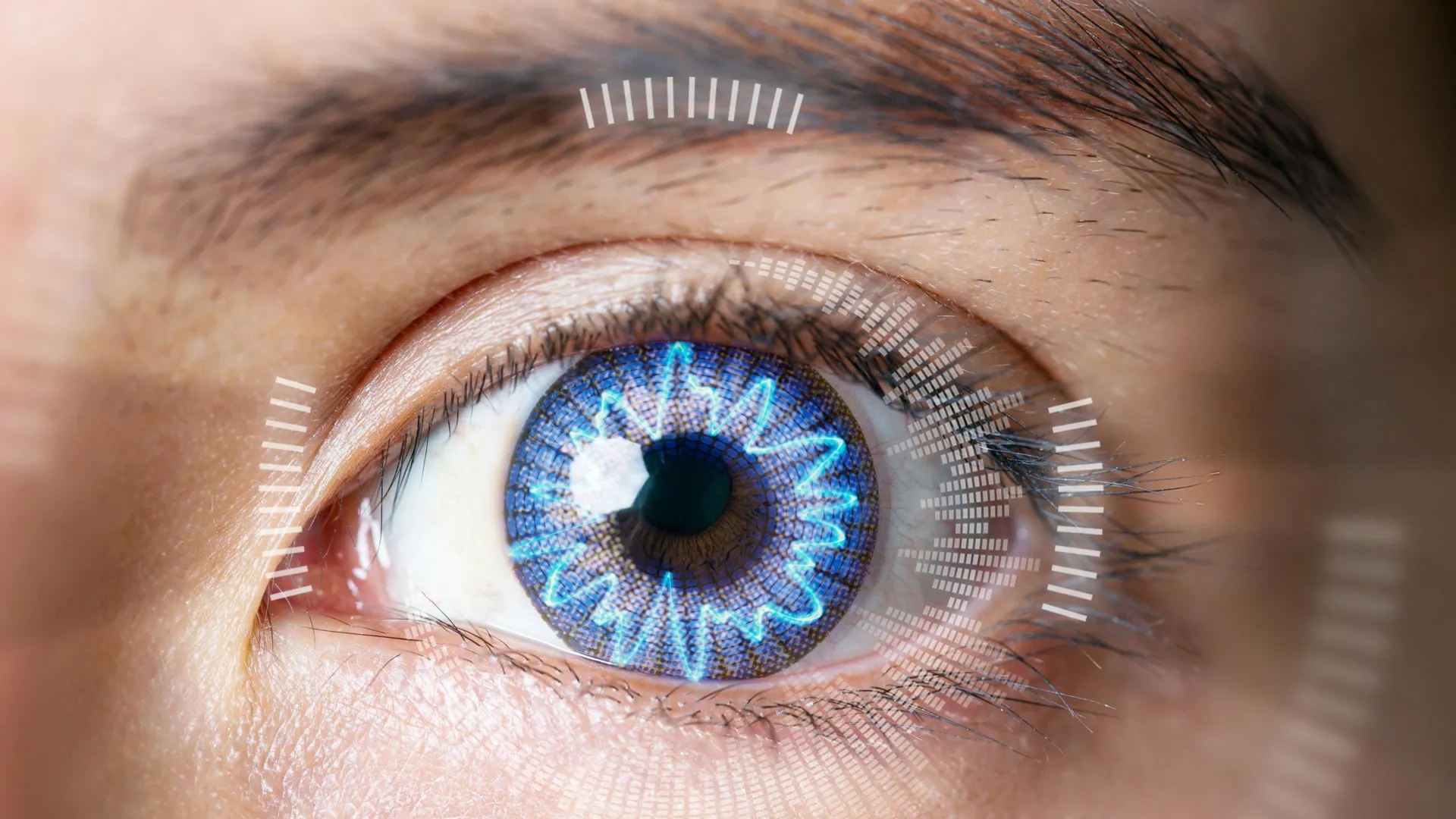
Millions of Americans have altered vision, ranging from blurriness to blindness. But not everyone wants to wear prescription glasses or contact lenses. Accordingly, hundreds of thousands of people undergo corrective eye surgery each year, including LASIK — a laser-assisted surgery that reshapes the cornea and corrects vision. The procedure can result in negative side effects, prompting researchers to take the laser out of LASIK by remodeling the cornea, rather than cutting it, in initial animal tissue tests.
Michael Hill, a professor of chemistry at Occidental College, will present his team’s results at the fall meeting of the American Chemical Society (ACS). ACS Fall 2025 is being held Aug. 17-21; it features about 9,000 presentations on a range of science topics.
Human corneas are dome-shaped, clear structures that sit at the front of the eye, bending light from surroundings and focusing it onto the retina, where it’s sent to the brain and interpreted as an image. But if the cornea is misshapen, it doesn’t focus light properly, resulting in a blurry image. With LASIK, specialized lasers reshape the cornea by removing precise sections of the tissue. This common procedure is considered safe, but it has some limitations and risks, and cutting the cornea compromises the structural integrity of the eye. Hill explains that “LASIK is just a fancy way of doing traditional surgery. It’s still carving tissue — it’s just carving with a laser.”
But what if the cornea could be reshaped without the need for any incisions?
This is what Hill and collaborator Brian Wong are exploring through a process known as electromechanical reshaping (EMR). “The whole effect was discovered by accident,” explains Wong, a professor and surgeon at the University of California, Irvine. “I was looking at living tissues as moldable materials and discovered this whole process of chemical modification.”
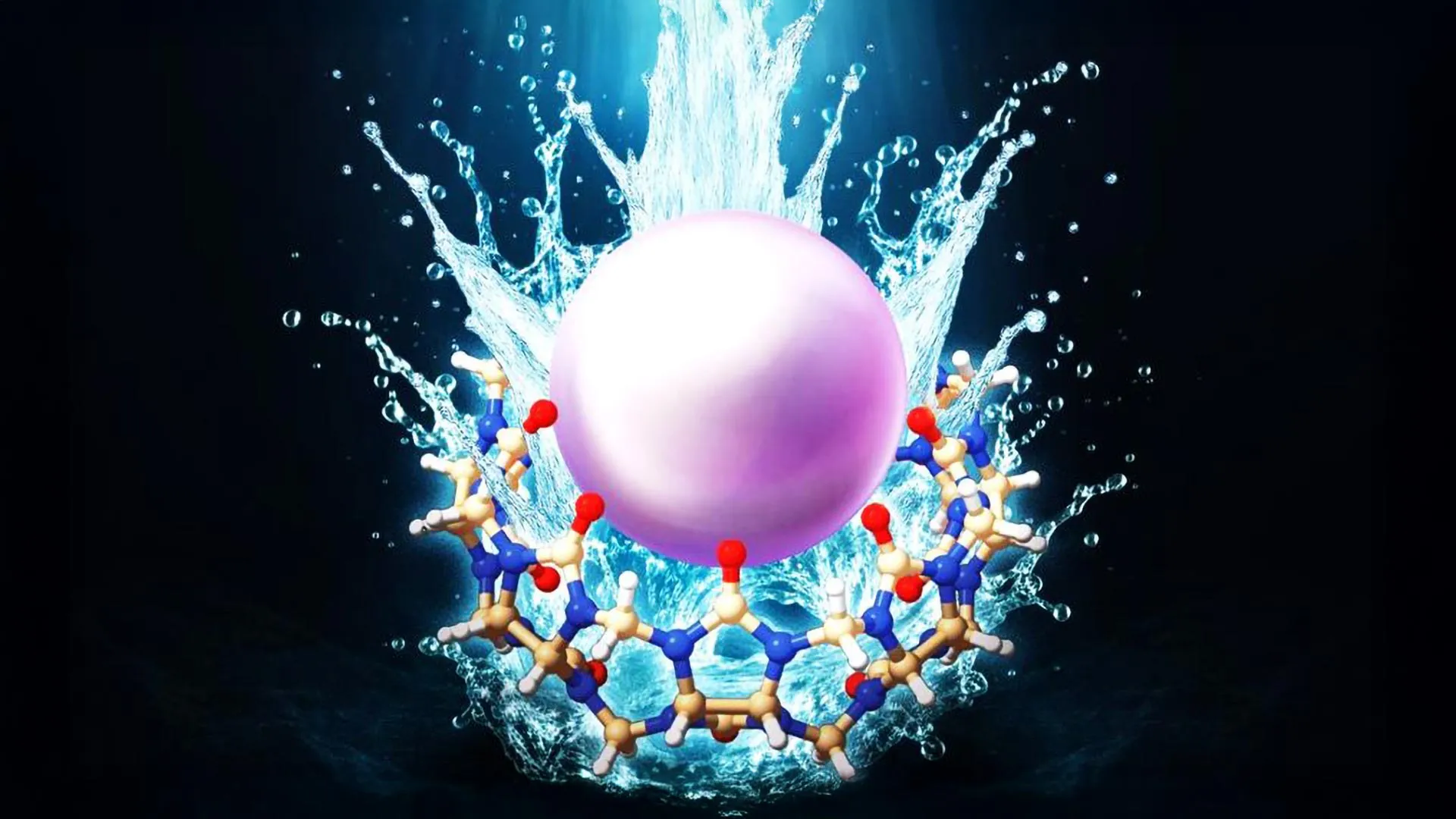
In the body, the shapes of many collagen-containing tissues, including corneas, are held in place by attractions of oppositely charged components. These tissues contain a lot of water, so applying an electric potential to them lowers the tissue’s pH, making it more acidic. By altering the pH, the rigid attractions within the tissue are loosened and make the shape malleable. When the original pH is restored, the tissue is locked into the new shape.
Previously, the researchers used EMR to reshape cartilage-rich rabbit ears, as well as alter scars and skin in pigs. But one collagen-rich tissue that they were eager to explore was the cornea.
In this work, the team constructed specialized, platinum “contact lenses” that provided a template for the corrected shape of the cornea, then placed each over a rabbit eyeball in a saline solution meant to mimic natural tears. The platinum lens acted as an electrode to generate a precise pH change when the researchers applied a small electric potential to the lens. After about a minute, the cornea’s curvature conformed to the shape of the lens — about the same amount of time LASIK takes, but with fewer steps, less expensive equipment and no incisions.
They repeated this setup on 12 separate rabbit eyeballs, 10 of which were treated as if they had myopia, or nearsightedness. In all the “myopic” eyeballs, the treatment dialed in the targeted focusing power of the eye, which would correspond to improved vision. The cells in the eyeball survived the treatment, because the researchers carefully controlled the pH gradient. Additionally, in other experiments, the team demonstrated that their technique might be able to reverse some chemical-caused cloudiness to the cornea — a condition that is currently only treatable through a complete corneal transplant.
Though this initial work is promising, the researchers emphasize that it is in its very early stages. Next up is what Wong describes as, “the long march through animal studies that are detailed and precise,” including tests on a living rabbit rather than just its eyeball. They also plan to determine the types of vision correction possible with EMR, such as near- and far-sightedness and astigmatism. Though the next steps are planned, uncertainties in the team’s scientific funding have put them on hold. “There’s a long road between what we’ve done and the clinic. But, if we get there, this technique is widely applicable, vastly cheaper and potentially even reversible,” concludes Hill.
Title Electrochemical corneal refraction
Abstract The cornea is a transparent, highly organized anatomical structure that is responsible for ~2/3 of the refractive power of the eye. The corneal stroma consists of orthogonally stacked collagen- fibril lamellae whose molecular composition and precise macromolecular geometry eliminate backscattered light and maintain the shape of the cornea. Anatomical variation, birth defects, trauma, and various pathologies can alter the shape, structural stability, and transparency of the cornea, thus affecting vision. Surgical interventions to treat myopia, hyperopia, and astigmatism include laser-assisted in situ keratomileusis (LASIK) and photorefractive keratectomy (PRK). Despite their popularity, these procedures are expensive and permanently lower the biomechanical strength of the cornea. Here we report our efforts to apply electromechanical reshaping (EMR) as a molecular- based, non-ablative/non-incisional alternative to laser vision refraction, using ex vivo rabbit globes. EMR relies on short electrochemical pulses to electrolyze interstitial water, with subsequent diffusion of protons into the extracellular matrix of collagenous tissues; protonation of immobilized anions within this matrix disrupts the ionic-bonding network that provides structural integrity. This leaves the tissue transiently responsive to mechanical remodeling; subsequent re-equilibration to physiological pH restores the ionic matrix, resulting in persistent shape change of the tissue. Optical coherence tomography (OCT), second-harmonic generation (SHG), and confocal microscopy suggest that EMR enables control over corneal contouring while maintaining the underlying macromolecular collagen structure and stromal cellular viability.
This research was funded by the National Eye Institute of the National Institutes of Health and the John Stauffer Charitable Trust.